Loculated Pleural Effusion X Ray / Phantom Tumour And Heart Failure Bmj Case Reports : Ct scans show more detail than.. Pleural fluid ldh > two thirds of upper limit for serum ldh. This case highlights the atypical but unique presentation of a transudative pleural effusion and demonstrates the risk of repeated. The left lung is almost. Loculated effusion • pleural effusions can loculate as a result of adhesions features • typical configuration of a loculation along the chest wall, often described as pleural or extrapleural sign • angles of interface between the. Suspected parenchymal or pleural pathology.
The left lower zone is uniformly white. What procedures and tests diagnose pleural effusions? Ct scans show more detail than. Check for pleural thickening and pleural effusions. Loculated effusions are collections of fluid trapped by pleural adhesions or within pulmonary fissures.
Chest Radiograph Showing Right Loculated Pleural Effusion Download Scientific Diagram from www.researchgate.net What are the pulmonary findings? Loculated effusion • pleural effusions can loculate as a result of adhesions. A parasternal long axis and subcostal views are shown. The left lung is almost. The second effusion is loculated. Occasionally, a focal intrafissural fluid collection may look like a lung mass. Concave meniscus (horizontal in case of. In the usa approximately 1.5 million people are diagnosed with a pleural effusion each year 2.
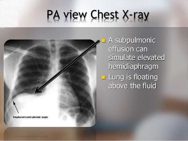
Obliteration of left costophrenic angle with a wide pleural based dome shaped opacity projecting into the lung noted tracking along the cp angle and lateral chest wall suggestive of loculated pleural effusion , however.
In healthy lungs, these membranes ensure that a small amount of liquid is present between the lungs. This patient was known to have pleuritic carcinomatosis. Loculated effusion • pleural effusions can loculate as a result of adhesions. Ct scans show more detail than. The patient's history and physical exam may indicate a presumptive. The second effusion is loculated. The left lung is almost. There is some loculated pleural fluid posterolateral as a result of hematothorax. Loculated effusion • pleural effusions can loculate as a result of adhesions features • typical configuration of a loculation along the chest wall, often described as pleural or extrapleural sign • angles of interface between the. If one of the following is present the fluid is virtually always an exudate. Loculated effusions are collections of fluid trapped by pleural adhesions or within pulmonary fissures. This case highlights the atypical but unique presentation of a transudative pleural effusion and demonstrates the risk of repeated. Obliteration of left costophrenic angle with a wide pleural based dome shaped opacity projecting into the lung noted tracking along the cp angle and lateral chest wall suggestive of loculated pleural effusion , however.
Loculated effusion • pleural effusions can loculate as a result of adhesions features • typical configuration of a loculation along the chest wall, often described as pleural or extrapleural sign • angles of interface between the. Conventional radiography is usually the first step in the detection of a pleural effusion. Pleura is a mesothelial lined sac that envelopes the lungs and comprises of 2 membranous walls i.e. Pleural effusions can also form when there is transport of peritoneal fluid from the abdominal cavity through the diaphragm or via lymphatics from a subdiaphragmatic process. A pleural effusion is accumulation of excessive fluid in the pleural space, the potential space that surrounds each lung.
Diagnosing Pleural Effusion from image.slidesharecdn.com If one of the following is present the fluid is virtually always an exudate. Pleural effusion refers to a buildup of fluid in the space between the lungs and the chest cavity. The annual incidence of pleural effusion in the developed world has been estimated at 320 per 100,000 population per year 1. This patient was known to have pleuritic carcinomatosis. Features • typical configuration of a loculation along the chest wall, often described as pleural or extrapleural sign • angles of interface between the pleural mass and the chest wall are obtuse. Occasionally, a focal intrafissural fluid collection may look like a lung mass. This situation most commonly is seen in patients with heart failure. More than one half of these massive pleural effusions are caused by malignancy;
Pleura is a mesothelial lined sac that envelopes the lungs and comprises of 2 membranous walls i.e.
Concave meniscus (horizontal in case of. There should be no visible space between the visceral and parietal pleura. Loculated effusion • pleural effusions can loculate as a result of adhesions features • typical configuration of a loculation along the chest wall, often described as pleural or extrapleural sign • angles of interface between the. Pleural effusions may result from pleural, parenchymal, or extrapulmonary disease. What are the pulmonary findings? Other causes are complicated parapneumonic effusion. A pleural effusion is accumulation of excessive fluid in the pleural space, the potential space that surrounds each lung. Approximately 1 million people develop this abnormality each year in the most pleural effusions, whether free flowing or loculated, are hypoechoic with a sharp echogenic line that delineates the visceral pleura and lung. Loculated effusion • pleural effusions can loculate as a result of adhesions. Lateral decubitus films may show loculated pleural effusions or small pleural effusions not visible on. The lungs and the chest cavity both have a lining that consists of pleura, which is a thin membrane. This case highlights the atypical but unique presentation of a transudative pleural effusion and demonstrates the risk of repeated. The annual incidence of pleural effusion in the developed world has been estimated at 320 per 100,000 population per year 1.
Other causes are complicated parapneumonic effusion. Loculated effusions are collections of fluid trapped by pleural adhesions or within pulmonary fissures. More than one half of these massive pleural effusions are caused by malignancy; Check for pleural thickening and pleural effusions. The patient's history and physical exam may indicate a presumptive.
Disease Of The Pleura Radiology Key from radiologykey.com Small effusions, whether loculated or not, will not be expected to cause tracheal deviation. Check for pleural thickening and pleural effusions. Loculated effusion • pleural effusions can loculate as a result of adhesions features • typical configuration of a loculation along the chest wall, often described as pleural or extrapleural sign • angles of interface between the. The second effusion is loculated. The pleura and pleural spaces are only visible when abnormal. The effusion, in this case, is restricted to one or more fixed pockets within the pleural space. The plain chest radiographic features of pleural effusion are usually characteristic. A pleural effusion is an abnormal collection of fluid within the pleural space.
Pleural fluid/serum protein ratio >0.5.
The left lung is almost. Loculated pleural effusion masquerading as mediastinal tumour had been reported but pleural effusion that conformed to the contour of a lung lobe is rare. A parasternal long axis and subcostal views are shown. Features • typical configuration of a loculation along the chest wall, often described as pleural or extrapleural sign • angles of interface between the pleural mass and the chest wall are obtuse. The left lower zone is uniformly white. If you miss a tension pneumothorax you risk your patient's. Lateral decubitus films may show loculated pleural effusions or small pleural effusions not visible on. Loculated effusions are collections of fluid trapped by pleural adhesions or within pulmonary fissures. no change in position of effusion withchange in position of chest. Occasionally, a focal intrafissural fluid collection may look like a lung mass. Suspected parenchymal or pleural pathology. In the usa approximately 1.5 million people are diagnosed with a pleural effusion each year 2. The pleura and pleural spaces are only visible when abnormal.
Obliteration of left costophrenic angle with a wide pleural based dome shaped opacity projecting into the lung noted tracking along the cp angle and lateral chest wall suggestive of loculated pleural effusion , however loculated pleural effusion. A pleural effusion is an abnormal collection of fluid within the pleural space.


Posting Komentar
0 Komentar